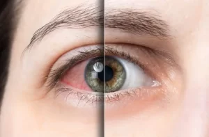
A retinal detachment refers to a serious eye condition where the light-sensitive tissue at the back of the eye separates from its underlying supportive layers. In this blog, we will explore the nature of a retinal detachment, its underlying causes, the telltale signs to watch out for, and the different treatment approaches available. Equip yourself with knowledge to take proactive steps in maintaining your eyesight. Now, let’s explore this condition and shed light on its intricacies.
What is retinal detachment?
A retinal detachment happens when the retina, the tissue at the back of the eye that detects light, separates from its usual position. It no longer sends visual information to the brain correctly. Act promptly if you notice symptoms like flashes, shadows, or blurred vision. These indicate a detachment occurring. See an eye doctor right away for an examination. Retinal reattachment procedures like laser therapy, freezing treatment, or surgery can repair the detachment before permanent vision loss occurs. The sooner the condition is treated, the more likely you can stabilise or partially restore vision. Contact your ophthalmologist urgently if you experience any worrying eye symptoms.
Symptoms of retinal detachment
Retinal detachment often brings sudden changes and vision problems. Early symptoms tend to escalate swiftly, so prompt medical attention is vital if you experience these troubling signs.
Flashes of light or sudden bursts

Sometimes, you may see flickers or flashes in your side vision. This could mean your retina is tearing.
A shadow or curtain in your peripheral vision
A warning sign of retinal detachment is noticing a shadowy area in your side vision. The spreading shadow seems like a veil blocking your view.
Blurry or distorted vision

Blurry vision or distorted images? This means your retina has detached from the back of your eye. This causes vision issues like wavy lines or blind spots.
Causes of retinal detachment
Catching retinal tears or detachment early is crucial for preserving vision and maximising treatment effectiveness. Several factors can lead to retinal separation:
Trauma or injury
Damage to the eye, whether a forceful strike or impact, can sever or separate the retina from its proper position. Even seemingly minor trauma during activities like bungee jumping, paintball, or airsoft can potentially lead to retinal detachment in some cases.
Ageing

As we age, the clear gel in our eyes may shrink and separate from the light-sensitive layer. This condition is posterior vitreous detachment. Sometimes, the shrinking gel can pull the retina out of place. In certain cases, the retracting gel exerts force, detaching the retina.
Other eye conditions
Eye issues may stress and detach the retina. Vision problems like nearsight, farsight, and irregular curves strain eyes. Cataracts and glaucoma raise risks. With diabetes, weak retinal vessels leak or bleed. Scar tissue then pulls the retina, causing detachment.
Family history and genetics
Retinal detachment sometimes runs in families. If a relative had it, there’s an increased risk. Certain genetic conditions like retinoschisis weaken the retina, raising detachment chances. A close relative’s history may signal a genetic predisposition.
Treatment options for retinal detachment
Picking a treatment depends on how bad the detachment is. Fast diagnosis and care boost the chances of stopping vision loss. See your eye doctor right away if you notice any symptoms.
Laser photocoagulation
Laser photocoagulation aims to seal retinal holes or tears. A laser emits light beams on areas around the tear. This helps bind the retina to underlying tissue, stopping fluid entry and detachment progression. The outpatient procedure takes place at the ophthalmologist’s office.
Cryopexy
Cryopexy freezes the retina to reseal tears. The doctor applies intense cold around the tear’s area. This creates scar tissue, securing the retina in place. Cryopexy treats tears where laser surgery isn’t viable due to the tear’s severity or location.
Scleral buckle surgery
Scleral buckle surgery treats severe retinal detachments. A flexible band wraps the eye’s white part, pushing the retina against the eye’s wall. This relieves fluid buildup under the retina. Freezing or laser seals holes or tears. This is an outpatient procedure, but recovery takes longer.
Pneumatic retinopexy
To fix a detached retina, air or gas is sometimes injected into the eye’s middle chamber. The bubble pushes the retina back against the eye wall. The gas gets absorbed, and scar tissue keeps the retina in place. This minimally invasive procedure works well for certain detachments.
Conclusion
Vision is precious, and the retinas are crucial to preserving it. A detached retina is a severe condition that demands prompt attention. If you experience sudden flashes, floaters, shadows, or vision impairment, seek medical assistance immediately. Early detection and treatment can prevent permanent vision loss. Laser surgery or cryopexy, when administered promptly, can often repair the issue and restore normal vision. Remain vigilant. You may also consider visiting Dr. JL Rohatgi Eye Hospital in Kanpur and consult their retina specialist for an efficient treatment if you notice any symptoms. They will examine your eyes, diagnose the problem, and recommend appropriate treatment options to prevent lasting damage. Remember, early intervention is key to treating retinal detachment successfully. Stay proactive and prioritise your eye health.


 Call Us Now
Leave a Reply